Aortic Coarctation
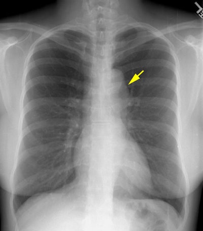
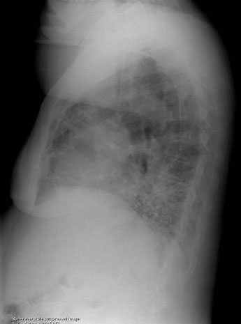
The 35 year old female patient in this case presented for routine chest radiographic evaluation.The plain film chest radiograph demonstrates a focal convexity to the descending aorta just distal to the aortic arch (yellow arrow). There is some prominence noted to the descending aorta just beyond the arch on the lateral view.
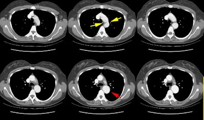
The CT scan demonstrated a focal aortic narrowing just beyond the ligamentum arteriosus (yellow arrows) with post-stenotic dilatation of the descending aorta (red arrow). An angiogram confirmed the CT findings and demonstrated a significant pressure gradient across the coarctation.
